If you or a loved one has been diagnosed with age-related macular degeneration (AMD), you've probably heard the terms "dry" and "wet" — and you may have wondered what they actually mean. They're not describing how your eye feels. They're describing two different disease processes happening in a tiny but critically important patch of tissue at the back of your eye. Understanding what's going on can make the diagnosis less mysterious and the treatment plan make a lot more sense.
First, Meet the Macula
The retina is the light-sensing tissue lining the back of the eye, working something like the film in an old camera. The macula is the small central portion of the retina — only about five millimeters across — responsible for your sharp, detailed, central vision. Reading, recognizing faces, driving, threading a needle: all of that depends on the macula. The rest of the retina handles your peripheral (side) vision, which is why even advanced macular degeneration does not cause total blindness. It affects the center of your vision, not the edges.
The macula is one of the most metabolically active tissues in the entire body. Its photoreceptor cells — the rods and cones that convert light into electrical signals — are constantly renewing themselves and consuming enormous amounts of oxygen and nutrients. All of that housekeeping depends on a support layer underneath the photoreceptors called the retinal pigment epithelium, or RPE. Think of the RPE as the macula's maintenance crew: it delivers nutrients, hauls away waste, and keeps the photoreceptors healthy. Beneath the RPE sits a thin membrane (Bruch's membrane) and, below that, a rich bed of blood vessels called the choroid that supplies the whole operation.
Macular degeneration, at its core, is a disease of this support system.
Dry AMD: A Slow Buildup of Waste
Dry AMD is by far the more common form, accounting for roughly 85 to 90 percent of cases. Here's the underlying process:
As we age, the RPE's waste-removal system becomes less efficient. Debris from the photoreceptors' constant renewal — a mixture of fats and proteins — begins to accumulate underneath the RPE, forming deposits called **drusen**. Your eye doctor can see drusen during an exam as small yellowish spots, and finding a few tiny ones is a normal part of aging. But when drusen become larger and more numerous, they signal that the maintenance system is falling behind.
The trouble is that drusen sit directly in the supply line. They act like a layer of sludge between the choroid's blood vessels and the RPE, making it harder for oxygen and nutrients to reach the cells that need them. Over years, chronically stressed RPE cells begin to sicken and die. And because photoreceptors depend completely on the RPE, when a patch of RPE dies, the photoreceptors above it die too.
In its advanced stage, dry AMD produces well-defined patches of lost RPE and photoreceptors called **geographic atrophy** — essentially, islands of retina that no longer function. When these patches involve the center of the macula, patients notice blurred or missing spots in their central vision. This process is typically slow, unfolding over years, which is why many people with dry AMD maintain good functional vision for a long time.
Inflammation also plays a starring role. Research has shown that an ancient part of the immune system called the complement system becomes chronically overactive in AMD, contributing to RPE damage. This discovery led to the first FDA-approved treatments for geographic atrophy, which work by dialing down complement activity.
Wet AMD: When Blood Vessels Break the Rules
Wet AMD is less common — about 10 to 15 percent of cases — but historically it has caused the majority of severe vision loss from this disease. Importantly, wet AMD is not a separate disease; it's a complication that develops on top of the dry process.
Here's how it happens. When the retina is starved of oxygen and stressed by inflammation, it sends out a chemical distress signal — most notably a molecule called **VEGF** (vascular endothelial growth factor). VEGF is essentially a message that says "grow new blood vessels here." In many parts of the body, that's a helpful repair mechanism. In the macula, it's a disaster.
In response to VEGF, abnormal new blood vessels sprout from the choroid and grow through breaks in Bruch's membrane into the space under the retina — a process called **choroidal neovascularization**. These new vessels are hastily built and fragile. They leak fluid, they leak fats, and they bleed. That's the "wet" in wet AMD: fluid and blood accumulating where they don't belong, lifting and distorting the delicate retinal layers.
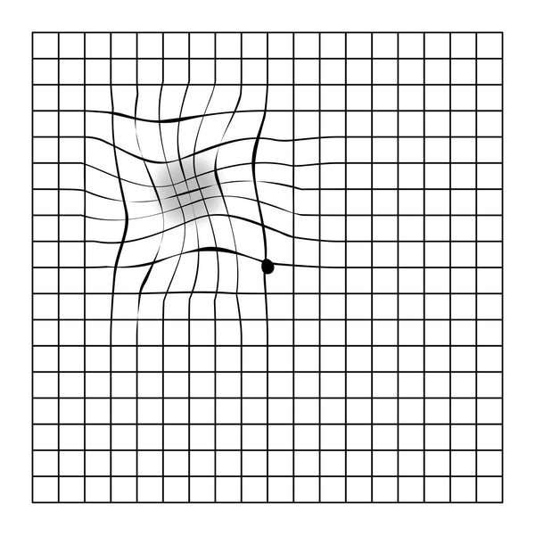
This is why wet AMD symptoms tend to appear suddenly. Straight lines look wavy or bent. A dark or blurry spot appears in central vision. Print becomes distorted rather than just dim. If leaking and bleeding continue unchecked, scar tissue forms in the macula, causing permanent central vision loss — sometimes over just weeks to months rather than years.
The silver lining: because we understand the VEGF signal driving this process, we can block it. Anti-VEGF medications, injected into the eye, neutralize the distress signal, causing the abnormal vessels to stop leaking and often regress. These treatments have transformed wet AMD from a condition that reliably caused legal blindness into one where most patients maintain — and many even regain — useful vision, provided treatment starts promptly.
Why the Distinction Matters
Dry AMD is a slow degeneration driven by waste accumulation, poor nutrient supply, and chronic inflammation. Wet AMD is an acute complication driven by abnormal, leaky blood vessel growth. Both stem from the same underlying breakdown of the macula's support system, and anyone with dry AMD carries some risk of converting to the wet form — which is why regular monitoring matters so much.
If you've been diagnosed with AMD, a few things are worth remembering. Check each eye individually at home (an Amsler grid — a simple grid of straight lines — makes distortion easy to spot), and call your retina specialist promptly if straight lines start to bend or a new blur appears in your central vision. With wet AMD, days matter. Nutritional supplementation based on the AREDS2 study, not smoking, and controlling cardiovascular risk factors all help slow progression. And keep your scheduled eye exams, even when your vision feels stable — the changes that matter most often start before you notice them.
Macular degeneration is a serious diagnosis, but it is no longer the untreatable condition it was a generation ago. Understanding the biology behind it is the first step in becoming an active partner in protecting your sight.
*This article is for general education and is not a substitute for a personal evaluation by an eye care professional.*